
Our Services
High quality X-Ray and ultrasound imaging tailored to your needs.
Your referral may use medical shorthand. This page translates the main examination groups into plain English and explains the usual preparation. If you are still unsure, send us the referral. We would rather check it than have you guess.
X-Ray
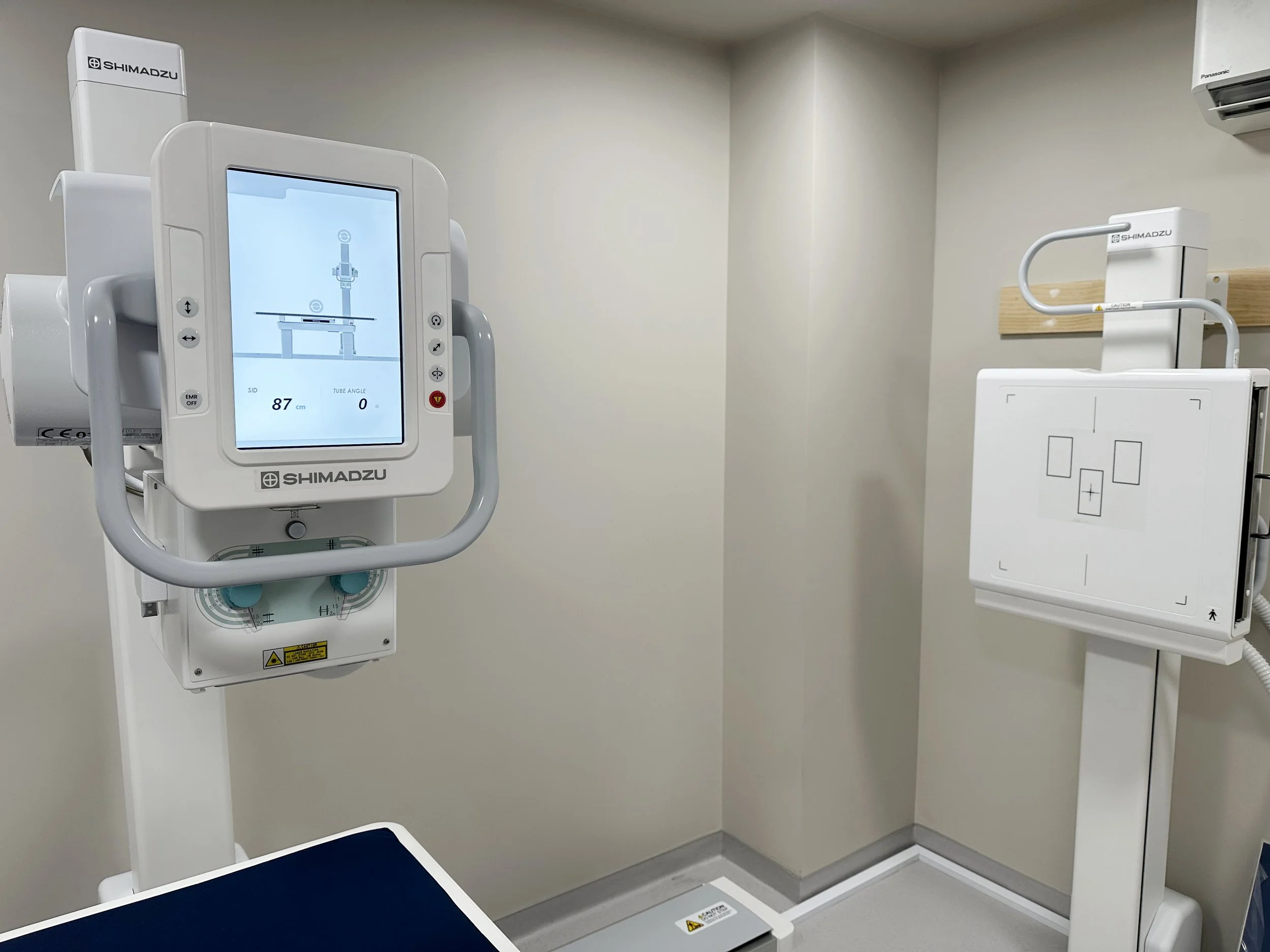
X-Ray is a fast imaging technique commonly used to assess bones, joints, the chest and other structures.
Common examinations: chest; selected abdominal imaging; cervical, thoracic and lumbar spine; pelvis and hips; upper and lower limbs; long length lower limb and spine imaging.
Preparation: Most general X-Rays require little preparation. You may be asked to remove jewellery, metal objects or clothing over the area. Please tell us if you are pregnant or think you may be pregnant.
Ultrasound
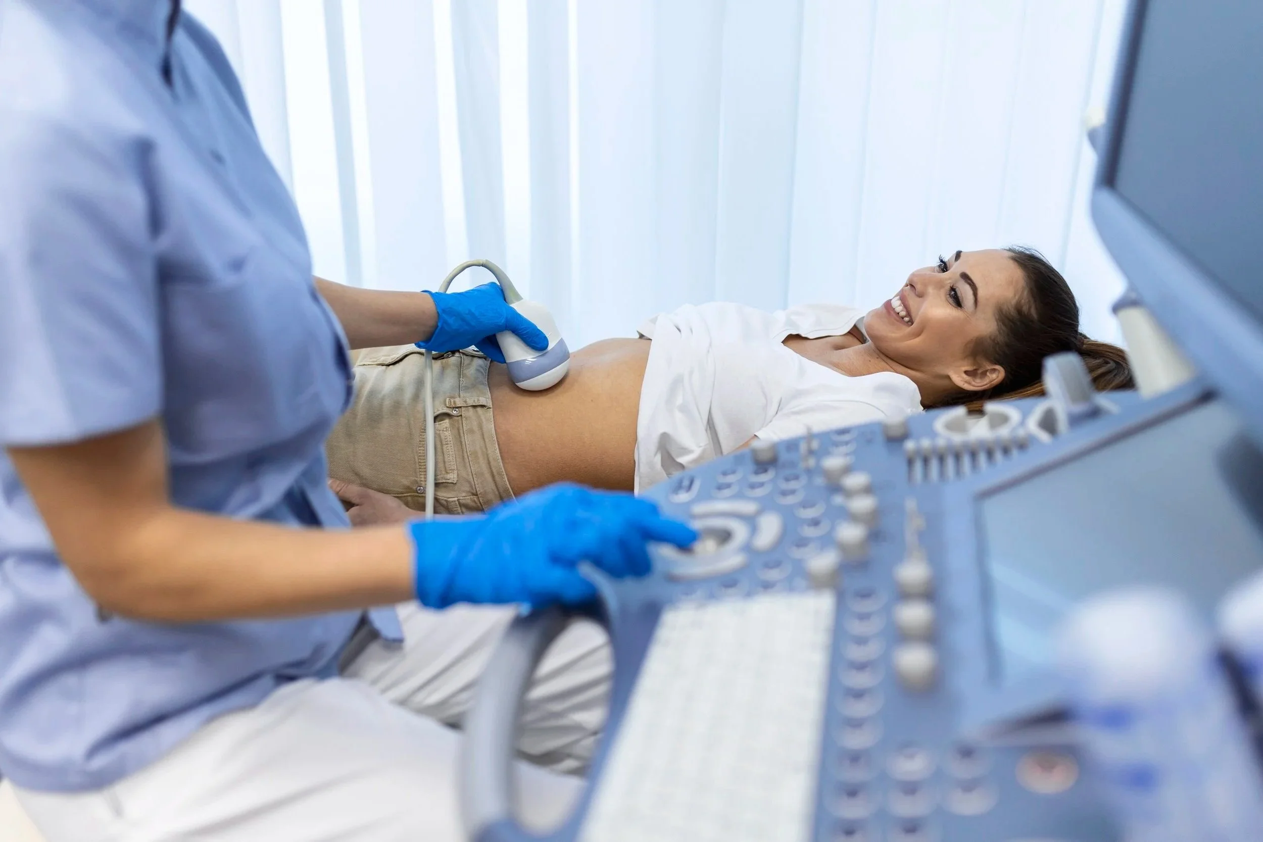
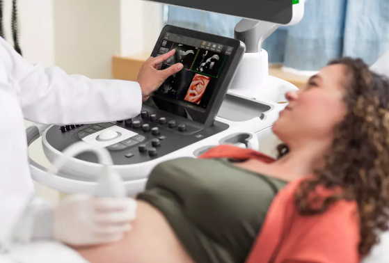
Ultrasound uses high frequency sound waves to create live images of organs, soft tissues and blood flow. It does not use X-Ray radiation.
Scans: abdomen; pelvis; renal and urinary tract; hernia; other general examinations reviewed by the clinical team.
Preparation: Abdominal ultrasound usually requires fasting for 6 hours. Pelvic ultrasound usually requires a comfortably full bladder. Combined abdomen/pelvis may require both.
Pregnancy Ultrasound
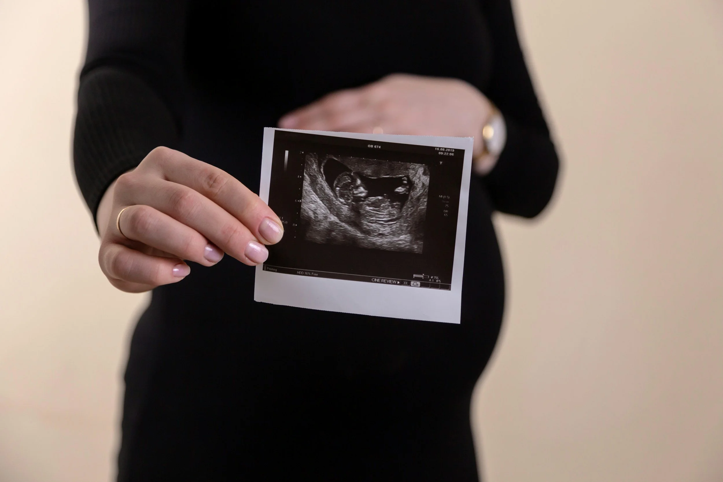
Specialist pregnancy ultrasound through different stages of pregnancy.
Scans: dating and viability; nuchal translucency screening; anatomy; growth; Doppler assessment when clinically requested.
Preparation: Preparation and timing vary by scan.
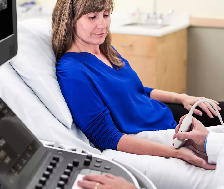
MSK Ultrasound
Real time assessment of muscles, tendons, ligaments, joints and selected peripheral nerves.
Common areas: shoulder, elbow, wrist, hand, hip, knee, ankle, foot and selected soft tissue regions.
Preparation: Usually no special preparation unless advised.
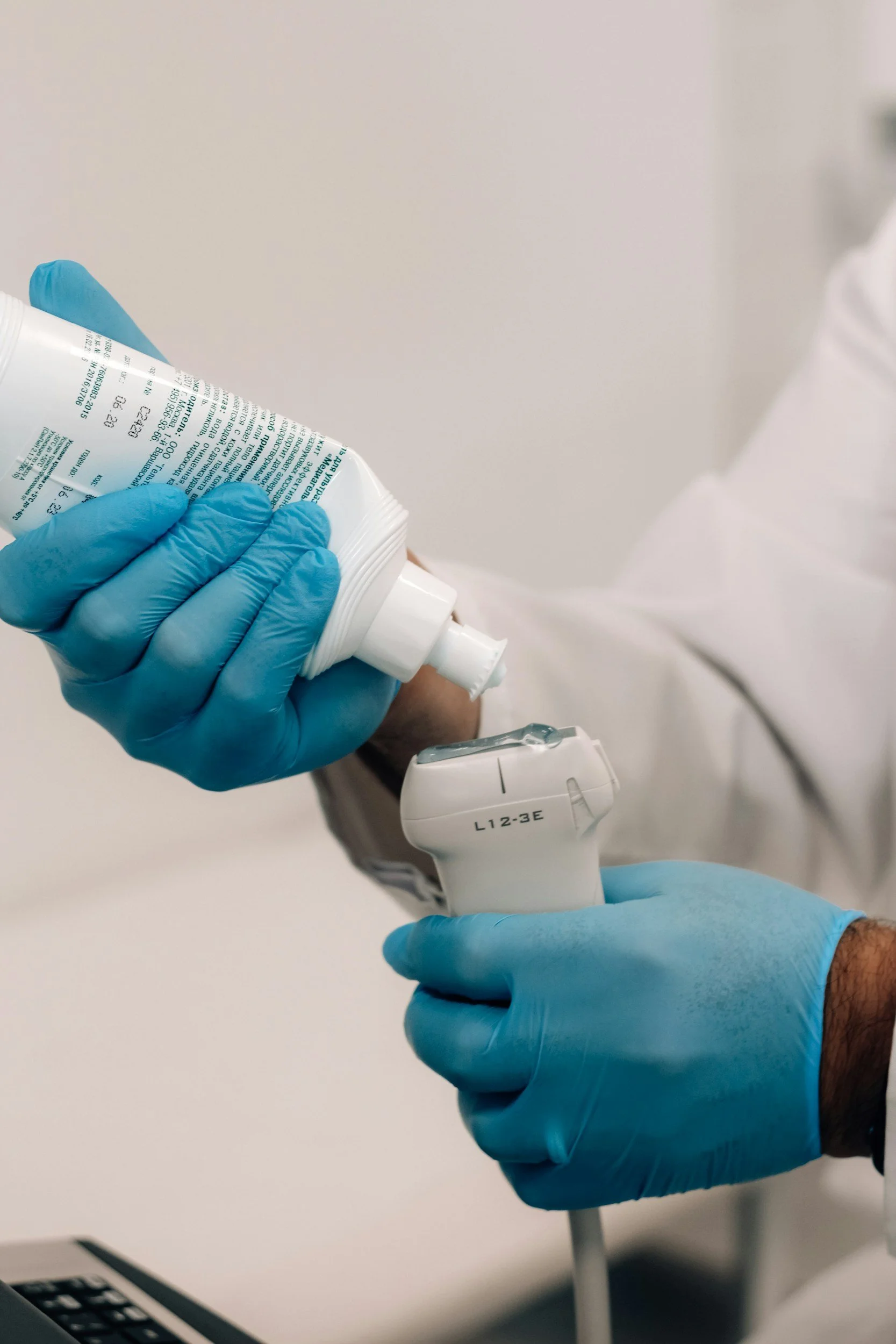
Small Parts Ultrasound
High resolution ultrasound for superficial structures.
Scans: thyroid; salivary glands; scrotal/testicular; groin; soft tissue lumps; foreign body localisation.
Preparation: Usually no special preparation unless advised.
Vascular Ultrasound
Specialised imaging to assess blood flow and selected vascular conditions.
Scans: upper limb DVT; lower limb DVT; venous mapping; abdominal aortic aneurysm surveillance; carotid arteries.
Preparation: Preparation depends on the examination. Final instructions should be confirmed by the clinical team.
Need help choosing the right scan?
Send us your referral or call us. Our team can check the request and organise the correct appointment.